Plasmon Interactions: Using Nanoparticles to Enhance Optical Phenomena
 Another of the directions of the research that my group and I have carried out is using arrays of noble metal nanoparticles to enhance optical processes due to interactions with particleplasmons. It’s interesting to note that Nanotechnology has been around in some sense for hundreds of years. Some of the brilliant colors of stained glass windows in cathedrals across Europe derive from the formation of nanoclusters of certain metals when added to glass. Gold clusters yield red glass, for example.
A much more contemporary application of the interaction of light and nanoparticles is in fabrication of biosensor arrays or biochips, which are often based upon fluorescence. The appeal of this is the ease with which biochemists can add a fluorescent tag to molecules which in turn bind to certain biomolecules of interest. This in fact is the basis of the so-called sandwich array. There has been a good deal of work done investigating a related technique, called surface enhanced Raman scattering, or SERS, which allows a different type of molecular fingerprinting. There huge enhancements, up to 10^14 have been reported. Much less work has gone into optimizing and understanding the enhancement which comes from the interaction of light from fluorescent molecules and metallic nanoparticles. Fluorescence of course involves both absorption and radiative decay, at two different wavelengths, due to the Franck-Condon Effect. Our results show evidence that the observed nanoparticles enhancement of fluorescence involves both processes.
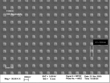
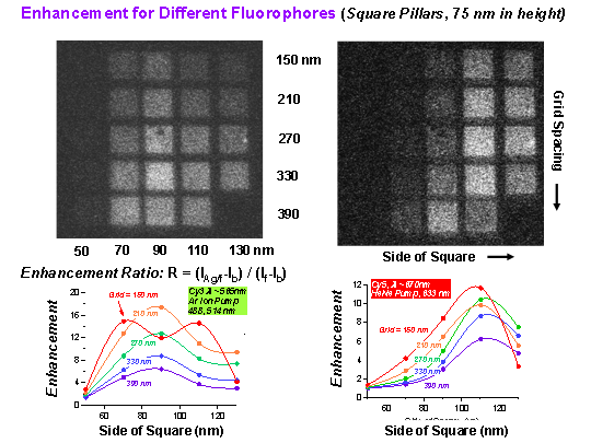 We use electron beam lithography to create arrays of silver nanoparticles on a substrate. This allows us control over the shape, size and spacing of the particles-all of which effect the plasmon resonance frequency. To avoid quenching of the fluorescence we need a finite spacing between the fluorescent molecule and the nanoparticles. We use a combination of proteins to space a variety of fluorescent tags several nm from the surface of the surface of the Ag particles. We measure the fluorescence using a scanning laser microscope and a filter which cuts out the incident light, but allows a band of wavelengths through to the detector. It allows us to quickly determine the optimum combination of size an spacing as seen in the figure to the right.. The upward shift in the optimum size in going from green light to red points to an enhanced absorption of the incident light, since the plasmon resonance is known to shift to lower frequency for larger particles. On the other hand, we find that the maximum in intensity coincides with a minimum in the fluorescence life time showing that the emission is also enhanced. Our present work is aimed toward a quantitative, and predictive understanding of nanoparticle-enhanced fluorescence, and related phenomena.